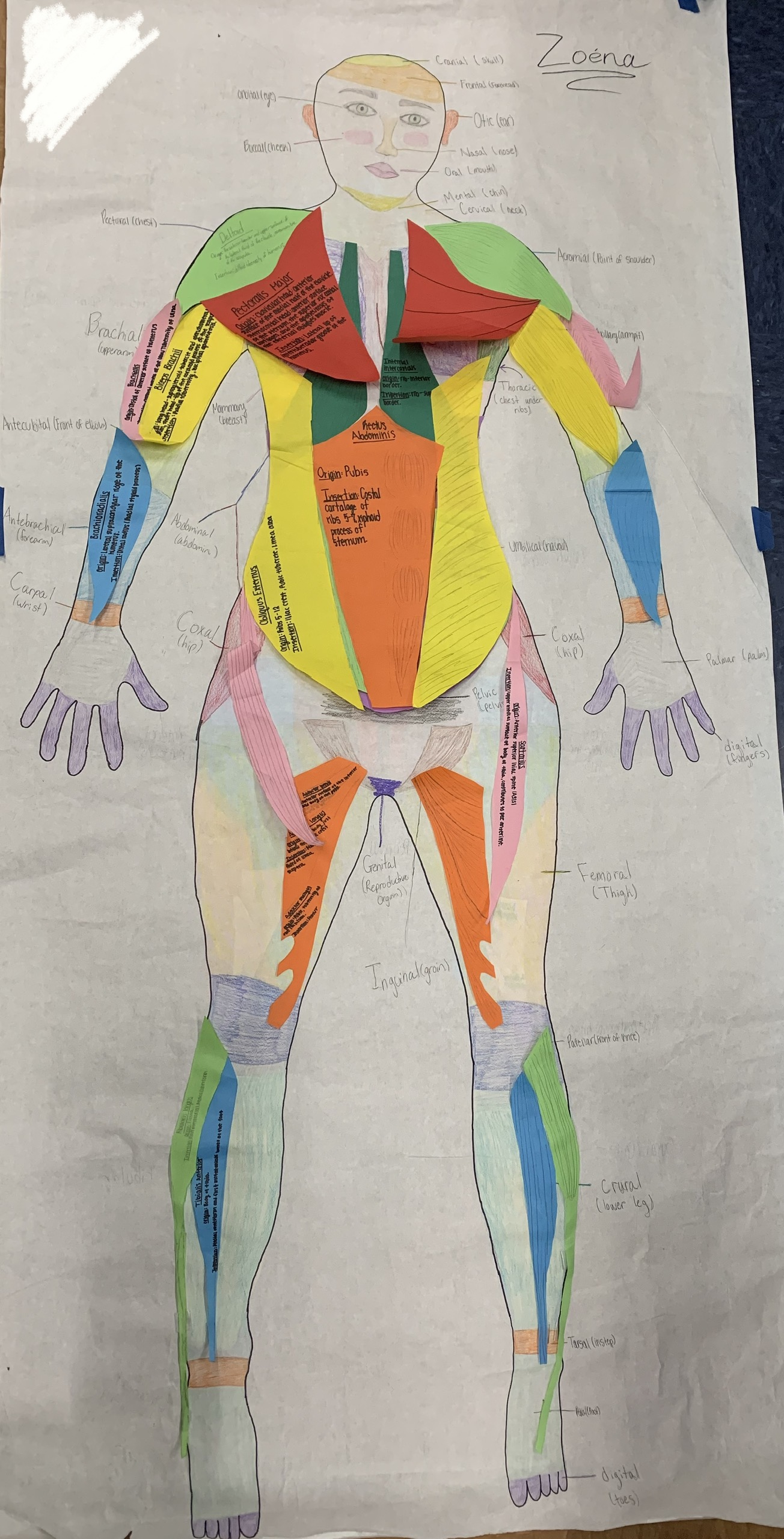
Students created life-size body drawings and added muscles.
Four of our 14 anatomy students plan a health-related career.
Another student plans to study forensics.
The remaining 9 have no concrete plans for what they will do in 5 months, after graduation.
We needed a helpful way to learn about muscles. Sure, we could use drawings and label them after a lecture, and memorize names, origins, insertions, actions, and innervation, quiz one another and then take quizzes and get grades.
Ugh.
Instead, we used our life-size human drawings to learn about muscles.
Our learning progressed like this:
- We had a short lecture on basic muscle structure.
- We learned how muscles were related to bones and movement by origin, action, and insertion.
- We learned the histology of 3 types of muscle cells.
- We did a series of station activities that reinforced our learning.
- We constructed life-size paper models of 42 muscles and labeled their action, origin, and insertion.
- We added the paper muscle models to our life-size drawings.
- Finally, each student selected a movement of some type from a list of movements I provided (or a movement of their own choosing) and made a presentation to each classmates about the muscles (and bones) involved in the movement.
The discussions as students figured out how best to represent muscles on their model blew me away. Students talked their way through deciding how best to label each muscle and how to represent muscles as the 3-D objects students had never actually seen, using only strips of paper. Considerations included how best to model the origin and insertion attachments, which muscles laid over or under others, and even individual differences from one group’s model to other models. Students wondered if human muscles actually actually had differences in size and shape from one person to another.
There were several styles of presentations. Some students made slide shows, some used their muscle models and short video clips to explain the movement they chose. Some physically demonstrated the movement themselves, inviting classmates to participate and feel their own muscles moving. One student made a clever video about their movement.
After experiencing al the presentations, students reflected on their learning. All students said they remembered names, actions, origins, and insertions of most of the muscles in their own presentation and could describe how the muscles worked together to perform the movement they demonstrated. They stated that they learned more about the structure and function of the bones involved. All said they knew how to find the information needed to understand muscles and that they’d be able to do the same set of steps for another movement using different muscles.
Will students remember all the names of all the muscles, actions, origins, and insertions years from now? Probably not. Did students gain a deeper understanding than if they’d painstakingly memorized muscle names, origins, insertions, and actions for a test? Most certainly. Did students connect and transfer their learning to new scenarios? Not totally yet – but this was a great start.
Our journey through connecting and transferring learning through collaboration:
- Part 1 of Learning Anatomy through Collaboration – Regions of the Body and Anatomical Terms
- Part 3 of Learning Anatomy through Collaboration – The Nervous System Introduction
- Part 4 of Learning Anatomy through Collaboration – Nerves, Muscles, and The Brain (coming soon)

[…] Part 2 of Learning Anatomy through Collaboration – Muscles […]
[…] systems, including tendons and ligaments.Some students chose the same scenario they used in Part 2 – muscles, and added the nervous […]
[…] Part 2 of Learning Anatomy through Collaboration – Muscles […]